

Illustrative consultation about three-dimensional dental imaging
General & Family Dentistry

CBCT 3D Dental Scans in Mississauga

“Scan” can mean several different things in a dental office. For people coming from Mississauga, here is a plain guide to CBCT 3D dental scans: how they differ from other imaging, when one might be suggested, and what happens to the results.

Sorting out the terms, Mississauga

Three things called a scan, and only one is CBCT

An optical impression scanner sweeps a small wand over the teeth and records their surface with light. A panoramic X-ray sweeps around the head and produces one wide, flat picture. Cone-beam computed tomography, CBCT, also uses X-rays, but it collects data that can be viewed in three dimensions, showing teeth, bone and neighbouring structures in depth. Knowing which of these was meant clears up a lot of confusion.

CBCT tends to come up when a particular question cannot be resolved by an exam and conventional images. Typical examples involve planning around nerves or sinuses, or a tooth problem that remains unexplained. A recommendation is easier to judge when it comes with the reason, the alternatives, what the scan may not reveal and the exposure involved.

Contact Milton Dental Clinic to go over an imaging suggestion. Have any referral ready, plus where and when your last X-rays were taken. Ask who would perform the scan, who reads it and how results reach your treating dentist. Submitting an online form does not confirm a scan, on-site imaging or that CBCT fits your case.

Clarify which scan was recommended
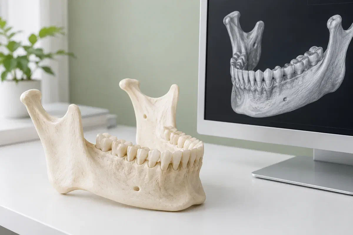
Illustrative jaw teaching model beside a generic three-dimensional dental rendering
Typical reasons CBCT comes up

When flat images leave a gap

Three broad questions account for many CBCT discussions. Having one of these concerns does not mean a scan is automatic; it means the option may be worth weighing.

Working near delicate anatomy

Implants and some surgical procedures sit close to nerves, sinuses and other roots. Depth information can clarify those relationships. Ask what exactly is uncertain, how it affects the proposal, and whether a different route would remain open.

A root or tooth problem still unexplained

After an exam and routine X-rays, some endodontic or other dental problems remain unclear. Extra imaging may narrow things down. It works alongside what you report and what is found clinically, and it cannot always deliver a firm diagnosis.

Locating something unusual

Teeth that have not come in, or unexpected features on another image, sometimes need a more precise sense of where they are or how far they extend. Ask why the suggested scan area is right for this. A larger field is not inherently more informative.

Asking “could we manage with the images we already have?” is always reasonable. A lower-dose X-ray may sometimes provide what is needed.

What CBCT can contribute

Depth, used sparingly

Unlike a single flat picture, CBCT data can be examined as thin slices and rotated to different viewpoints. In the hands of a qualified clinician and for the right question, that may assist with diagnosis, planning or reviewing an area over time. Its value always sits on a scale opposite the exposure.

Ionizing radiation is part of every CBCT exam, and the dose is generally greater than for routine dental X-rays. Equipment, settings and the size of the region all influence it. Good practice limits the scan to what the question requires while keeping the images diagnostically useful. That is a long way from claiming zero risk or quoting one universal number.

Patient information: FDA — dental cone-beam computed tomography

A reason you can repeat back

After the explanation, you should be able to say what the scan will help decide. If the main selling point is that the equipment is advanced, ask again what clinical choice depends on it.

Context for every finding

Interpretation draws on your history, the exam and earlier records. Ask who will read the scan and how they will handle anything noticed outside the original question.

Records that can move between offices

Where a separate imaging provider is involved, find out how images and the report will be transferred, with your permission, so the treating clinician can actually use them at your next appointment.

What to expect

Recommendation, preparation, scan, review

Imaging and treatment discussions can be booked separately. Check the provider, the address, the fee and the reporting plan before setting out from Mississauga, especially when the request came from another dentist.

  1. Confirm the purpose

    The clinician responsible looks at why 3D imaging is being considered and whether images on file already cover it. Give the dates and places of any earlier scans. Ask what the scan is expected to add.

  2. Tell the team what they should know

    Follow the preparation you are given. Mention pregnancy or the possibility of it, trouble staying still, or any need for positioning support. Earrings, glasses, hairpins or removable dental appliances may need to come out or off.

  3. The scan itself

    You are positioned, usually seated or standing, and asked to stay still while the device moves around your head. Details vary by machine and by the area imaged. If you feel uncomfortable, say so before it starts.

  4. Results, explained

    Someone qualified must interpret the images and prepare findings. Ask when they will be available and who will walk you through them. You may be offered further assessment or a changed plan depending on what is seen.

Following up

Where does the report go from here?

Taking the images is only the collecting part. They become useful through interpretation and follow-up, which can need extra attention when the scan happened somewhere other than your dental office.

Find out who is calling

Check whether you should arrange a results appointment yourself or expect a call from the referring office. If a procedure is booked around the report, confirm its arrival before taking the plan as final.

Keep the essentials written down

Note the scan date, the provider and the area covered. If new imaging is proposed in future, mention this exam first. It may or may not answer a later question, but looking at it can prevent a needless repeat.

Symptoms still need attention

A pending or completed scan is not a reason to wait out increasing pain, new swelling or another fresh problem. Let the treating team know if things change while you wait, so they can judge how soon to see you.

Choosing among imaging options

Which view fits the question?

Imaging should be picked to answer the clinical question, no more and no less. Ask whether the benefit justifies a CBCT and whether another approach would work.

When the exam and records cover it

No new imaging

Sometimes your history, a thorough exam and current images give enough to decide. A treatment conversation does not by itself mean a new scan. Ask the clinician to be open about what is still uncertain.

When a single plane shows enough

A standard dental X-ray

Bitewing, periapical or panoramic images can sometimes answer the question. Each has a different role from CBCT. Ask why a particular view is proposed and whether its detail is enough.

When depth makes the difference

A targeted CBCT

The provider selects an exam suited to the question and arranges how it will be reported. Coverage and settings should reflect what must be seen and your individual factors. Larger or sharper is not, by itself, a justification.

Imaging terms, answered

Questions about the different scans

Bring the note or referral that mentions a scan. Looking at the wording together often clears up which exam is meant and who should answer each practical detail.

(905) 876-4488
How is a panoramic X-ray different from a CBCT?
A panoramic X-ray produces a single wide, flat image of the teeth and jaws. CBCT collects data that can be viewed in three dimensions and in slices. They answer different questions, and the exposure differs as well, so ask which one is proposed and why.
My dentist used a wand to scan my teeth. Was that a CBCT?
Probably not. A handheld intraoral scanner records tooth surfaces with light to create a digital impression. CBCT is an X-ray exam of internal structures that uses a larger unit positioned around the head.
Does a 3D scan use more radiation than my usual check-up X-rays?
Generally, yes. CBCT usually involves more exposure than routine dental X-rays, though dose varies with the machine, settings and area covered. Your dentist should explain why the expected benefit justifies it in your case.
Can a CBCT see inside my sinuses or jaw joint?
Depending on the area scanned, CBCT can include parts of the jaw and nearby structures. Whether that is relevant depends on the clinical question. Ask what the scan is intended to examine, and who will interpret areas outside the teeth.
Do I need to arrange my own scan before seeing a dentist?
Usually not. It is generally better to be assessed first so the clinician can decide whether a scan is needed and of what area. Bring details of earlier imaging instead of booking a scan independently.
Who reads the scan if it is taken at a separate imaging centre?
Confirm this ahead of time. The scan needs proper interpretation and a report sent to the clinician managing your care. Ask who will contact you, when, and how any further recommendations will be followed up.

Patient information: RCDSO — dental CT scanner requirements

CBCT questions from Mississauga

Not sure which scan you were offered?

Send the details to our Bronte Street practice and we can help sort out what was recommended and how it relates to your dental care. We will point you toward the next step. Please confirm the imaging provider and your appointment time before going for any scan.

This form starts an enquiry; it does not book an imaging appointment. Phone us if a sore or swollen area needs quick attention.

Visit Milton Dental Clinic
Unit 1, 342 Bronte St. S, Milton, ON L9T 5B7
Get directions Clinic hours
Monday–Thursday
11am–7pm
Friday–Sunday
By appointment only

Areas we serve

  • Milton
  • Oakville
  • Mississauga
  • Georgetown
  • Halton Hills
  • Acton
  • Campbellville
  • Brampton
  • Burlington
  • Puslinch
  • Rockwood
  • Erin
  • Caledon
  • Etobicoke
  • Old Milton
  • Bronte Meadows
  • Timberlea
  • Dorset Park
  • Dempsey
  • Clarke
  • Beaty
  • Coates
  • Harrison
  • Scott
  • Willmott
  • Ford
  • Bowes
  • Cobden
  • Walker
  • Mountain View
  • Milton Heights
  • Hawthorne Village
  • Valley View
  • Fallingbrook
  • Nassagaweya
  • Trafalgar
  • Esquesing
  • Nelson
  • Brookville
  • Moffat
  • Glen Williams
  • Norval
  • Limehouse
  • Stewarttown
  • Ashgrove
  • Hornby
  • Silver Creek
  • Speyside
  • Scotch Block
  • North Oakville
  • River Oaks
  • Glen Abbey
  • West Oak Trails
  • Palermo
  • Bronte Creek
  • College Park
  • Joshua Creek
  • Uptown Core
  • Preserve
  • Iroquois Ridge
  • Lisgar
  • Meadowvale
  • Meadowvale Village
  • Churchill Meadows
  • Erin Mills
  • Central Erin Mills
  • Streetsville
  • East Credit
  • Creditview
  • Heartland
  • Sheridan
  • Mount Pleasant
  • Credit Valley
  • Huttonville
  • Churchville
  • Springbrook
  • Northwest Brampton
  • Bram West
  • Fletcher's Meadow
  • Fletcher's Creek South
  • Alton Village
  • Orchard
  • Millcroft
  • Headon Forest
  • Tansley
  • Brant Hills
  • Tyandaga
  • Kilbride
  • Lowville
  • Aberfoyle
  • Morriston
  • Arkell
  • Hillsburgh
  • Freelton
  • Carlisle
  • Waterdown
  • Flamborough
  • Mountsberg

How can we help?

Milton Dental Clinic

Have a question about your smile? Choose the option that works best for you.

For urgent dental concerns, please call.
Messages are reviewed during clinic hours.