

Illustrative consultation about three-dimensional dental imaging
General & Family Dentistry

CBCT 3D Dental Scans in Old Milton

Years of dental records already tell a story. In Old Milton, a conversation about CBCT 3D dental scans starts with what those images show, what they leave unanswered, and whether a three-dimensional view would actually change your care.

Old Milton and the long view

Your history comes first, the scanner second

Plenty of people around Milton’s historic downtown have seen the same teeth through many check-ups, fillings and perhaps a crown or two. That record is valuable. When cone-beam computed tomography (CBCT) is mentioned, the first step is usually to ask what the existing exam notes and X-rays already settle, and what they cannot.

CBCT uses X-rays to gather three-dimensional information about the teeth, jaws and nearby structures. It is meant for selected questions, like the shape of bone at a site being considered for an implant, that a flat image leaves unclear. It should not be confused with the camera-style scanner some offices use to capture tooth surfaces for a digital impression. A recommendation deserves a stated reason, along with the alternatives, the limits of what a scan can show, and the radiation it involves.

Call or write to Milton Dental Clinic if you would like to talk a recommendation through. Mention any referral, and list where earlier X-rays or scans were taken. Ask who would perform the scan, who would read it, and how the report would reach your treating dentist. An online message is an enquiry only and does not confirm imaging or its suitability.

Talk through an imaging question
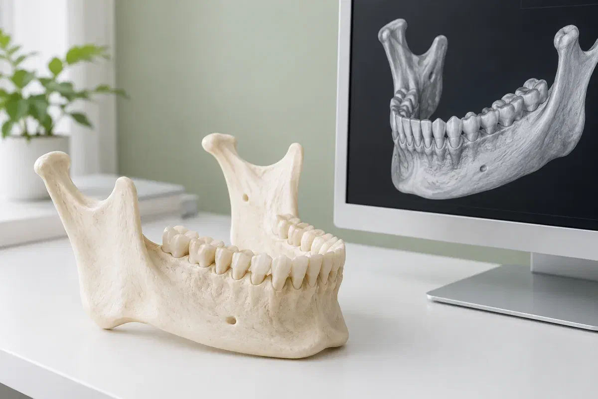
Illustrative jaw teaching model beside a generic three-dimensional dental rendering
Questions a 3D view can help with

When the old films stop short

The situations below are reasons a scan might be discussed, not signs that one is required. Your dentist should connect any recommendation to your own findings.

Bone and anatomy before a procedure

Replacing a long-missing tooth with an implant, or planning certain oral surgery, can depend on bone volume and where nerves or sinuses sit. Ask which detail is unclear, how the answer would steer the plan, and whether a non-surgical choice is still open.

An older tooth with a puzzling problem

A tooth with years of work behind it may develop a problem that standard images do not explain, sometimes after root canal treatment. A scan may add information. Your symptoms and the exam remain central, and the image will not always give a firm answer.

A shadow that needs a second look

An unerupted tooth or another unusual finding may need its position or extent clarified. Ask why the proposed scan area suits the question. Capturing more of the jaw is not automatically better when the concern is small and specific.

CBCT is not a regular part of a check-up. Ask whether images already on file, or a lower-dose X-ray, could give the information needed.

Where 3D information earns its place

A view in slices, read by someone who knows your mouth

With a three-dimensional data set, a qualified clinician can move through an area layer by layer and from different directions. When the scan is well chosen, that can support a diagnosis, a treatment plan or a follow-up evaluation. Those gains only count if they outweigh the exposure for the particular question.

Dental CBCT uses ionizing radiation, and the dose is usually higher than for routine dental X-rays. It changes with the unit, its settings and the size of the area imaged. A sensible scan is limited to what the question requires while still giving enough detail to be useful. Nobody can fairly promise no risk, or give one dose figure that applies to everyone.

Patient information: FDA — dental cone-beam computed tomography

Purpose stated up front

Ask which choice depends on the result. A helpful answer ties the scan to something concrete, like understanding the anatomy before a procedure, rather than describing the machine as modern.

Read alongside your records

Your history, the exam and earlier images all inform how a scan is read. Ask who interprets it and how they will let you know about anything found outside the original question.

Shared with permission

If an imaging provider takes the scan and another clinician treats you, ask how the images and report will be passed along with your authorization, so the next visit is not spent waiting for files.

Step by step

What happens between the suggestion and the answer?

The scan and the treatment discussion may not happen on the same day or in the same building. Before going, confirm the provider, the location, the fee and how the report will be delivered.

  1. Review the reason and the archive

    Your dentist considers why a scan is being suggested and whether earlier images settle the matter. Share dates and places of past scans if you know them. Ask what new information is expected and how it could change what is planned.

  2. Get ready, and speak up

    Follow the provider’s instructions. Mention if you are or could be pregnant, if staying still is hard, or if you need help getting into position. Hearing aids, dentures, glasses or jewellery near the area may need to come off.

  3. Have the scan taken

    The team positions you and tells you how to stay still while the equipment circles your head. What this looks like depends on the unit and the area. Mention any discomfort before the scan begins so adjustments can be considered.

  4. Hear what it shows

    Images need careful interpretation by a qualified person. Confirm when results will be ready and who will explain them. Depending on the findings, further assessment or a different treatment approach may be suggested.

Afterward

Close the loop on the result.

A scan collects information and nothing more. It becomes useful through a proper reading and follow-through, especially if the imaging took place away from the dentist managing your care.

Ask who calls whom

Check whether you should book a visit to discuss results or wait for the referring dentist to reach out. If treatment depends on the report, make sure it has been received before you assume the plan is set.

Add it to your own file

Jot down when and where the scan was done and which area it covered. If another clinician proposes imaging later, mention this scan. It may not be enough on its own, but reviewing it first can avoid repeating an exam unnecessarily.

Do not wait on symptoms

An upcoming or finished scan is no substitute for prompt care if pain worsens, swelling appears or something new develops. Let the treating team know about any change while results are pending so they can reassess the timing.

Three levels of imaging

Start with what you have.

The right imaging is the one that answers the clinical question with no more exposure than needed. Ask whether the benefit justifies a scan, and whether another method could do the job.

When the record already answers it

Exam plus existing images

Your history, a careful exam and recent images may be enough to decide. Discussing treatment does not by itself mean a new scan is needed. Your dentist should be clear about any uncertainty that remains.

When a two-dimensional view will do

A conventional X-ray

An intraoral or panoramic image may supply what is required. It serves a different purpose from CBCT. Ask why a certain view is suggested and whether it gives enough detail for the planned assessment.

When depth is truly required

CBCT, when justified

A qualified provider selects the scan and how it will be reported. The area covered and the settings should match the diagnostic need and your own factors. Availability of a larger or sharper image is never a reason in itself.

Old Milton CBCT questions

What long-time patients tend to ask

Bring the referral or written recommendation with you. Talking it through can clarify what is being requested and whether your dentist or the imaging provider is best placed to answer each practical point.

(905) 876-4488
My X-rays go back many years. Could they make a new scan unnecessary?
Sometimes. Older images can show how an area has changed over time, and recent ones may answer the current question. Your dentist will judge whether they are adequate. Let the team know where earlier images were taken so they can be requested if useful.
I have several crowns and fillings. Will they affect a CBCT scan?
Metal and some dental materials can create streaks or shadows on CBCT images, which may limit what can be seen in nearby areas. Your dentist and the imaging provider take this into account when deciding whether a scan is likely to help.
Do I need to remove my partial denture for the scan?
You may be asked to take out removable appliances, along with jewellery, glasses or hearing aids near the area being scanned. Follow the provider’s instructions, and ask beforehand if you are unsure what to remove.
How is a dental CBCT different from an impression scan?
A CBCT uses X-rays to look at structures inside the teeth and jaw. An impression scanner uses light to record the outer shape of teeth, often for a crown or appliance. Check which one has been recommended, since the reasons and preparation differ.
Is the dose from a CBCT scan the same for everyone?
No. It depends on the equipment, the settings and how large an area is captured. CBCT generally involves more exposure than standard dental X-rays. Ask how the scan will be limited to what your question needs, and what the expected benefit is.
If the scan finds something unrelated, who tells me?
Ask this before the scan. The person interpreting the images should report relevant findings to the clinician managing your care. Clarify who will contact you, roughly when, and how any further recommendations will be followed up.

Patient information: RCDSO — dental CT scanner requirements

CBCT conversations in Old Milton

Bring the paperwork; we will help sort the next step.

Contact our Bronte Street practice with your questions and any referral or imaging details. We can explain how a scan might fit into your dental assessment and what would come next. Confirm the imaging provider and appointment details before attending for a scan.

Online requests are enquiries, not confirmed imaging appointments. A sore or puffy area that needs a quick look is better handled by phone.

Visit Milton Dental Clinic
Unit 1, 342 Bronte St. S, Milton, ON L9T 5B7
Get directions Clinic hours
Monday–Thursday
11am–7pm
Friday–Sunday
By appointment only

Areas we serve

  • Milton
  • Oakville
  • Mississauga
  • Georgetown
  • Halton Hills
  • Acton
  • Campbellville
  • Brampton
  • Burlington
  • Puslinch
  • Rockwood
  • Erin
  • Caledon
  • Etobicoke
  • Old Milton
  • Bronte Meadows
  • Timberlea
  • Dorset Park
  • Dempsey
  • Clarke
  • Beaty
  • Coates
  • Harrison
  • Scott
  • Willmott
  • Ford
  • Bowes
  • Cobden
  • Walker
  • Mountain View
  • Milton Heights
  • Hawthorne Village
  • Valley View
  • Fallingbrook
  • Nassagaweya
  • Trafalgar
  • Esquesing
  • Nelson
  • Brookville
  • Moffat
  • Glen Williams
  • Norval
  • Limehouse
  • Stewarttown
  • Ashgrove
  • Hornby
  • Silver Creek
  • Speyside
  • Scotch Block
  • North Oakville
  • River Oaks
  • Glen Abbey
  • West Oak Trails
  • Palermo
  • Bronte Creek
  • College Park
  • Joshua Creek
  • Uptown Core
  • Preserve
  • Iroquois Ridge
  • Lisgar
  • Meadowvale
  • Meadowvale Village
  • Churchill Meadows
  • Erin Mills
  • Central Erin Mills
  • Streetsville
  • East Credit
  • Creditview
  • Heartland
  • Sheridan
  • Mount Pleasant
  • Credit Valley
  • Huttonville
  • Churchville
  • Springbrook
  • Northwest Brampton
  • Bram West
  • Fletcher's Meadow
  • Fletcher's Creek South
  • Alton Village
  • Orchard
  • Millcroft
  • Headon Forest
  • Tansley
  • Brant Hills
  • Tyandaga
  • Kilbride
  • Lowville
  • Aberfoyle
  • Morriston
  • Arkell
  • Hillsburgh
  • Freelton
  • Carlisle
  • Waterdown
  • Flamborough
  • Mountsberg

How can we help?

Milton Dental Clinic

Have a question about your smile? Choose the option that works best for you.

For urgent dental concerns, please call.
Messages are reviewed during clinic hours.