Clarifying anatomy for a procedure
Implant placement and some surgery depend on bone and on where nerves or sinuses lie. Ask which relationship needs clarifying, how the report will be used in planning, and what alternatives exist.


A 3D scan is only as good as the reading behind it. From Puslinch, learn who interprets a CBCT 3D dental scan, how incidental findings are handled, why imaging may be recommended in the first place, and how the report reaches your dentist.
A cone-beam computed tomography (CBCT) scan produces a large set of three-dimensional X-ray data covering teeth, jawbone and whatever else falls inside the scanned area. That data has to be reviewed carefully by a qualified person, not just the tooth that prompted the scan. Knowing who that person is, and how their conclusions will reach you, is as important as the appointment itself.
A scan is considered when a specific question needs depth that an exam and conventional X-rays have not provided, such as anatomy near a planned implant or a dental problem without a clear cause. It is unrelated to the optical scanner used to take digital impressions. Along with the reason, you should hear about the alternatives, the limits of what imaging can show and the radiation it involves.
Contact Milton Dental Clinic with questions about a scan recommendation or report. Have any referral ready, and note where earlier images were taken. Ask where the scan would be performed, who would read it and how a written report would come back to your dentist. Reaching out online starts a discussion only; it does not secure an imaging slot or settle whether CBCT is warranted.
These are situations in which CBCT is sometimes discussed. They are reasons for a conversation, not a list of conditions that always require imaging.
Implant placement and some surgery depend on bone and on where nerves or sinuses lie. Ask which relationship needs clarifying, how the report will be used in planning, and what alternatives exist.
Some root canal or other dental issues are not fully explained by routine images. A scan may help, though it is interpreted together with your symptoms and the exam, and the report may not offer a definitive answer.
A tooth that has not erupted, or an unusual area on another image, may need its position or extent described more precisely. Ask why the scan area suits the question, since a wider field also means more anatomy to interpret.
CBCT is not a routine check-up image. Ask whether existing X-rays, or a simpler lower-exposure view, could answer the question first.
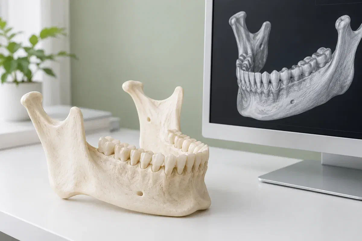
Because CBCT data can be viewed in thin sections and rotated, a trained reader can follow structures in ways a flat image does not allow. When the scan suits the question, that interpretation may support diagnosis, treatment planning or later evaluation. The benefit has to justify the exposure.
The exam uses ionizing radiation and usually delivers more than routine dental X-rays. Equipment, settings and the size of the scanned area all affect the dose. Good practice keeps the volume as small as the question allows while producing images adequate for a confident reading. It is not risk-free, and no single dose describes every scan.
Patient information: FDA — dental cone-beam computed tomography
Ask what the clinician will do with the findings. A clear purpose, such as checking anatomy before a procedure, helps the reader focus and helps you understand the result.
Your history, the exam and earlier records frame how the data is reviewed. Ask who is responsible for reviewing everything in the scanned area and how findings beyond the original question will be communicated.
If the imaging provider and treating clinician differ, ask how the images and written findings will be shared, with your authorization, so your next appointment can make use of them.
Different people may handle each stage, sometimes in different places. Confirm the provider, the location, the fee and the reporting arrangement before setting out from Puslinch.
The responsible clinician considers why imaging is needed and whether earlier images already answer the question. Share details of previous scans. Ask what the report is expected to clarify.
Follow the imaging provider’s instructions. Mention a possible pregnancy, trouble staying still or any need for positioning help. You may need to remove jewellery, glasses or removable dental work.
A technologist lines up your head, then the scanner arm makes its sweep while you hold steady. The routine depends on the machine and area. Raise any discomfort before the scan begins.
The images are interpreted and the findings documented. Ask when the report will be ready and which clinician will go through it with you. The results may lead to further assessment or a change in plan.
The scan only gathers information. Its value comes through interpretation and follow-up, which can stall if the images were taken somewhere other than the office managing your care.
Find out whether you should book a results appointment or expect a call. If treatment is waiting on the findings, confirm the report has reached your dentist before assuming the plan is ready.
Keep the date, provider and area scanned, and ask whether you can have a copy of the report. If new imaging is proposed later, share this one so it can be reviewed first.
A pending or completed report does not replace prompt assessment of worsening pain, swelling or a new problem. Tell the treating team about changes so they can reconsider timing.
Bring your referral and any earlier reports. Reviewing them together helps clarify what was requested and whether your dentist or the imaging provider should answer specific questions.
Patient information: RCDSO — dental CT scanner requirements
Send our Bronte Street team any referral or earlier report, along with what you would like explained. We can explain how a scan might fit into your dental assessment and what follow-up to expect. Confirm the imaging provider and appointment before attending.
Online requests are enquiries, not booked scans. If you have pain or swelling that needs attention soon, phone the office.
