Documenting a visible chip or worn surface
An image can record where an edge has changed. It does not confirm depth or cause. Share any sensitivity, grinding or past injury so the record reflects the whole picture, not just appearance.


Once a photo of your tooth is taken, it is fair to wonder what happens to it. Intraoral camera images help explain visible findings during the exam, and they may also become part of your dental record, so it is worth knowing how they are used and what they can show.
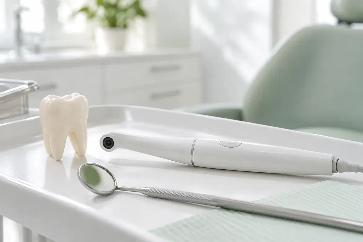
During the visit, the purpose is simple: to help you see what your dentist sees. An intraoral camera is a lightweight, pen-shaped instrument that takes magnified photographs of tooth and gum surfaces within reach. The image appears on a screen, where a worn surface, a filling edge or a hard-to-clean area can be pointed out directly.
After that, the picture may be kept with your clinical notes where appropriate. Either way, it remains a photograph, shaped by light, moisture, angle and what the lens could reach. Its meaning comes from the dentist’s interpretation alongside your symptoms, history and the rest of the examination, not from the image alone.
From Acton, northwest of Milton, you can ask when booking whether photos would help with your concern and how the visit can be set up. You are also welcome to ask how images are stored, who may see them and what consent applies to sharing. In the chair, ask which tooth is shown, which detail matters and what is still unclear.
A photo should serve a clear purpose. Mention the area you want explained and ask how the picture relates to the full exam.
An image can record where an edge has changed. It does not confirm depth or cause. Share any sensitivity, grinding or past injury so the record reflects the whole picture, not just appearance.
A close-up of a filling margin or a food trap can help explain a recommendation. A dark line still needs interpretation. Ask what other findings support any suggestion to replace the filling.
If a gumline area is hard to see at home, a photo can show where it is. Ask for a demonstration too, so the record leads to a change in your routine rather than sitting in a file.
Camera photographs, dental X-rays and 3D digital impressions are different records. They answer different questions and cannot replace each other.
In the moment, the photo lets your dentist indicate the exact feature while describing why it matters. That is especially helpful for back teeth and inner surfaces. You do not need to decode the image yourself; ask which area is important.
Later, a stored photo may support a review. Comparing images across visits can help, but differences in lighting and angle can change how a tooth looks. Any comparison should be read with a new exam and your history, rather than treated as proof by itself.
Patient information: Carestream Dental — intraoral camera image capture and review
Ask which tooth and surface the image shows. That helps you understand your notes, describe symptoms later and clean the right spot. Enlarged views can look unfamiliar, so ask how the image is oriented.
With the image in view, you can ask what problem a proposed treatment is meant to solve and what evidence beyond the photo points toward it. Alternatives, including waiting, may be available. A picture should help start a discussion of benefits and limits, not replace one, and you can take time to think when the situation is not urgent.
If an area is being followed, ask what will be compared next time. Photos can be useful with notes, measurements and symptoms. Ask which changes would warrant calling before the next review.
The camera supports a clinical visit. Mention tender areas, a strong gag reflex or limited opening so images can be captured with your comfort in mind.
The dentist examines the area and explains what a photograph would add. Ask the purpose. Some concerns cannot be answered visually, and a camera does not replace a proper assessment.
The small lens is placed near the relevant tooth or gum. Repositioning or blotting saliva might be needed for clarity. Use your agreed signal if you need a break.
While the image is on screen, ask which detail is significant and whether it is an observation or a diagnosis. A photo can show something is visible without proving depth, activity or cause. What goes into your notes should reflect the full assessment, including your symptoms and any tests, rather than the picture alone.
The visit may end with advice, monitoring, more tests or treatment options. Confirm the reason and timing. If images are kept or shared for your care, such as with another provider, ask how that works and what consent is needed.
Photos do not usually require any recovery care. The important part is following the plan based on the complete exam.
Before you leave, make sure you could describe the finding to a family member: the tooth involved, the likely explanation and anything still unresolved. Ask about any unfamiliar terms that may appear in your notes. A magnified surface can look dramatic, so the dentist’s explanation is what gives the image meaning.
Follow the instructions given for the specific issue found, whether it involves cleaning, a worn tooth or the gums. Ask to be shown a technique if useful. A stored photo does not repair a tooth or replace planned treatment.
If the area is being watched, note when it will be checked again and which symptoms mean calling sooner. Having a photo on file does not make future exams unnecessary. New pain or a change in function should be assessed promptly.
Privacy and paperwork questions are just as valid as clinical ones. Reading the photo is the dentist’s responsibility, and so is explaining how it sits in your file, what remains uncertain about the finding and what happens next.
Contact our Bronte Street practice with the concern you would like examined. Ask whether camera photos would add anything and how your visit can be planned. Your dentist will explain what the pictures show and what they cannot settle, and answer any questions about how they are kept.
Camera images support a clinical assessment. This page shows illustrative photography, not the clinic’s own equipment.
