A fresh chip or a worn corner
A close-up may show the outline of the missing piece or a flattened edge. Depth and cause need more than a photo. Tell the dentist about cold sensitivity, pain on biting, clenching or the moment you first noticed it.


You bite into a crust and suddenly your tongue finds a sharp new edge. An intraoral camera can photograph the chipped or worn surface up close, so you can see the change your dentist is assessing and talk through what it does and does not tell you.
A tongue is very good at finding new rough patches and very bad at telling you what they are. It might be a tiny chip, a worn corner, the edge of an old filling or a bit of hardened buildup. An intraoral camera, a compact handheld lens with a built-in light, can photograph the reachable surface so the dentist can show you which of these it actually is.
Still, a photo only captures what is visible at that angle, in that light, on a surface that may be wet or partly hidden. It cannot reveal how far a chip extends inside the tooth. Your dentist combines the image with what you have felt, your bite, your history and the rest of the exam before explaining what it means.
Coming down from Georgetown, you can ask when you call whether photographs would help with the tooth you have noticed and how the appointment can be organised. At the visit, ask which tooth is being shown, which part of the edge matters and what is still uncertain. Seeing a chip enlarged can be unsettling, so ask for perspective as well as a picture.
Images are most useful when tied to a specific question. Point to the tooth your tongue keeps finding and ask how the photo connects with other findings.
A close-up may show the outline of the missing piece or a flattened edge. Depth and cause need more than a photo. Tell the dentist about cold sensitivity, pain on biting, clenching or the moment you first noticed it.
Sometimes the sharp spot is the margin of an existing restoration. A photo can show this, but whether it needs smoothing, repair or nothing at all depends on the wider assessment. Ask what will decide that.
Hardened deposits along the gumline can feel like a new edge. An image can show where they sit, and the team can pair it with practical cleaning advice so the area is easier to keep clear at home.
Photographs from the camera are neither X-rays nor 3D scans. Each kind of record looks at a different question, so they are not interchangeable.
On screen, the dentist can trace the outline of a chip or a worn area while explaining whether it matters. That is often reassuring, because many rough spots turn out to be small. You do not need to analyse every highlight; ask which part of the picture shows the edge you have been feeling.
Where appropriate, the photograph can be added to your record. If the area is being watched, a later image may be compared with this one, keeping in mind that lighting and angle make a difference. A comparison should be read with a fresh exam and your history, not taken as proof of change by itself.
Patient information: Carestream Dental — intraoral camera image capture and review
Have the tooth and surface named for you. Knowing exactly where the edge is helps you describe any later symptoms accurately. Enlarged images can look odd at first, so ask how big the chip really is.
If smoothing, bonding or another repair is suggested, the image lets you ask what it would fix and what supports the recommendation. Leaving a small, stable chip alone may also be an option. Ask about benefits, limits and alternatives.
For a minor chip being watched, ask what will be checked next time and which signs would bring you back sooner. Photos can be useful alongside notes and symptoms.
The camera is used within a clinical visit. If the chipped tooth is sensitive, or you gag easily or struggle to open wide, mention it so the photographs can be taken gently.
The dentist asks about the moment you felt the edge, any pain, and your bite, then examines the tooth. Ask whether a photo would add anything. Some chips are clear without one, and the camera never replaces an assessment.
The lens is brought close to the chipped or worn area. The team may need a different angle or a quick blot of saliva for a clear image. Raise your hand if the tooth feels sensitive and you want them to stop.
Ask the dentist to outline the change and to separate what is visible from what is diagnosed. A photo cannot show how deep a chip runs or whether a crack lies beneath. Tests or other findings may fill in those gaps.
Options may include smoothing, a repair, monitoring or further tests. Confirm the reasoning and any timing. If the photo is kept in your record or shared for care, you may ask about storage and consent.
Being photographed needs no recovery care. What matters is the advice that follows from the full exam.
Before leaving, confirm whether it was a chip, wear, a filling margin or buildup, and what that means. Ask about any unfamiliar terms. A magnified chip can look bigger than it is, so ask for its real size.
You may be told to avoid biting hard foods on that side, adjust cleaning or protect the tooth in some other way. Ask for specifics. The photo does not repair the edge or replace any planned treatment.
If the chip is being monitored, note the next review and which symptoms mean calling earlier. A stored image does not replace a future exam. New pain, a tooth that feels loose or a piece breaking off should be checked promptly.
You do not need to interpret dental photos. The dentist should link the image to the exam, explain what is uncertain and answer questions about any next step.
Call our Bronte Street practice and describe the chip or rough spot you have noticed and when it appeared. The team can tell you if a camera view is likely to be useful and how to set up the visit. Your dentist will go through what the image shows, what it cannot establish and what can be done.
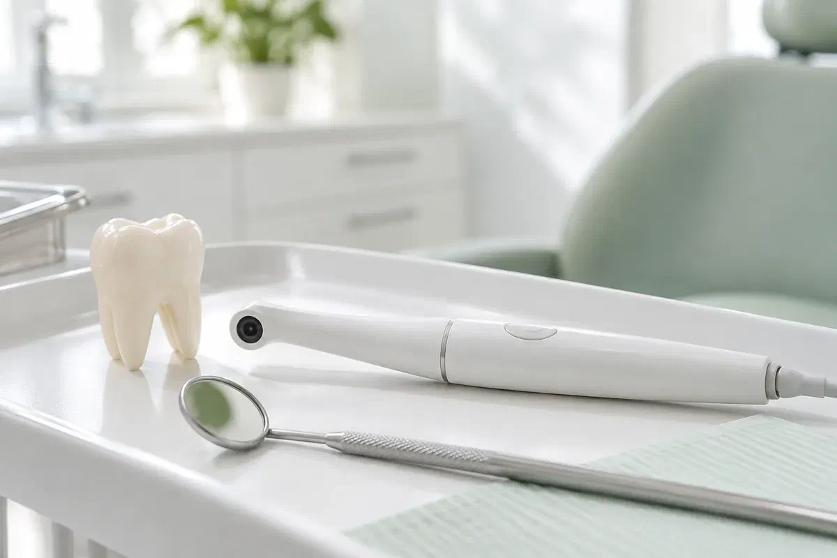
Photographs support a clinical assessment. Images on this page are illustrations, not pictures of the clinic’s equipment.
