Mild wear on a biting surface
A photo can show where enamel has flattened or chipped. It cannot measure depth or confirm why it is happening. Mention grinding, sensitivity or any bite change so the dentist can decide whether watching is appropriate.


“Let’s keep an eye on it” is common advice, but what exactly is being watched? An intraoral camera can photograph the visible surface in question, giving you a clearer idea of the spot under review and what would prompt a different plan.
Not every finding calls for treatment straight away. A small area of wear, a stained groove or an older filling edge may simply be monitored. That approach works best when you understand the spot itself, and an intraoral camera can help. The slim device photographs reachable tooth and gum surfaces at close range, so the dentist can show you what will be reviewed.
A picture is only one input. Lighting, saliva, the chosen angle and the parts of a tooth the lens cannot reach all limit what it shows. Before calling something stable or changing, the dentist weighs the photo against your symptoms, earlier records and the hands-on exam.
Households across Halton Hills, from Georgetown and Acton to Glen Williams and the hamlets in between, can ask when booking whether photographs suit the concern and how the visit can be arranged. In the chair, ask which tooth is displayed, which feature is being tracked and what would change the plan. A close-up should make watchful waiting feel informed, not uneasy.
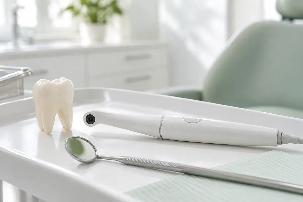
A photograph helps most when it records a specific concern. Tell the dentist which area you are wondering about and how it fits into the wider exam.
A photo can show where enamel has flattened or chipped. It cannot measure depth or confirm why it is happening. Mention grinding, sensitivity or any bite change so the dentist can decide whether watching is appropriate.
Some restoration margins look stained or uneven yet cause no trouble. An image helps you see what is being followed over time. Ask what signs would lead to more investigation or replacement.
If a gumline spot is being monitored, a photo can show where it is. Pair that with a cleaning demonstration so your home routine supports the review rather than leaving it to chance.
Camera photos are not dental X-rays and not three-dimensional scans. Each record answers a different question, so a monitoring plan may use more than one.
Seeing the area on screen lets the dentist point out exactly what is being followed and why. For a molar or a surface facing the tongue, that turns a vague note into something you can picture. Ask which part of the image is relevant instead of trying to judge every reflection.
Photos may be kept in your record where appropriate, which allows a later image to be set beside an earlier one. That comparison is helpful but imperfect: a slightly different angle, more light or a wetter surface can make a tooth look different. Changes should be judged with the exam and your history, not from two pictures alone.
Patient information: Carestream Dental — intraoral camera image capture and review
Ask for the tooth and surface to be named. That helps you notice relevant symptoms and clean the area well between visits. Magnified views can be disorienting, so ask how the image is oriented and how big the area really is.
Ask what change would move the plan from monitoring to treatment and which findings would support that. Other options may be available. A photo should help frame that conversation, not replace it.
Find out when the area will be looked at again and what will be compared. Photos can sit with measurements, notes and symptoms. Ask, as well, which signs mean contacting the clinic before the scheduled check.
Photographs are a supporting step inside a clinical appointment. Tell the team about tender spots, gagging or difficulty opening so images can be taken comfortably.
The dentist reviews your history and examines the area. Ask whether a photo will help with this review. Some changes cannot be judged by appearance, and imaging never replaces a proper assessment.
The camera is held close to the tooth or gum surface being followed. To keep the new image as comparable as possible with any earlier one, the team might try a similar angle and blot away saliva first. Signal if you need to pause, close your mouth or rest your jaw for a moment.
If earlier photos exist, the dentist may look at them alongside the new one, accounting for differences in light and angle. Ask what counts as an observation and what counts as a conclusion. A photo shows visibility, not depth, activity or cause.
The decision may be to continue watching, add tests, adjust home care or discuss treatment. Ask for the reasoning and the next review date. You can also ask how photos are stored and whether consent is needed to share them.
Being photographed needs no recovery care. Between appointments, what matters is following the advice and noticing changes.
Before leaving, confirm what was seen, what it suggests and which symptoms would be meaningful. Ask about any unclear terms. A magnified image can make a minor feature look worrying, so the explanation matters most.
Follow the cleaning or habit advice given for that specific finding, whether it is a brushing angle, a flossing aid or a night-time grinding concern. Ask for a demonstration if needed. Photographs do not protect a tooth; daily care and planned follow-up do.
A photo on file is not a substitute for a future exam. Keep the agreed review, and call earlier if pain begins, a tooth feels different when you bite or something breaks, even if the last photo looked fine.
You are not expected to interpret dental images yourself. The dentist should relate the photo to the exam, explain uncertainty and answer questions about any next step.
Contact our Bronte Street practice and mention the tooth or area that has been under review. Ask whether close-up photos would help and how the appointment can be set up. Expect a plain account of what each picture reveals, what it cannot tell us, and the kind of change that would move the plan from watching to doing something more.
Photos support a clinical assessment. Images on this page are illustrative and are not of clinic equipment.
