

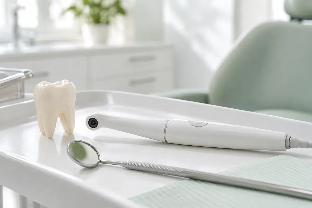
Illustrative dental consultation with a small intraoral camera, not actual clinic equipment
General & Family Dentistry

Intraoral Camera Examinations in Oakville

Being told a tooth needs work is easier to weigh when you can see what prompted the advice. An intraoral camera captures magnified photos of visible surfaces, so the reasoning behind a recommendation is laid out in front of you rather than taken on faith.

For decisions made from Oakville

What exactly is the dentist recommending, and why?

A treatment suggestion tends to raise two questions at once: what is wrong, and how sure are you? Seeing the area helps with the first. An intraoral camera is a compact, wand-shaped camera that photographs the reachable surfaces of teeth and gums up close, turning a verbal description into an image you can study with your dentist.

That image informs the exam but does not stand in for it. How a tooth appears on screen depends on lighting, angle, moisture and how much of the surface the lens can reach. The dentist still has to relate the photo to your symptoms, history and other findings before deciding what it means and whether treatment is truly needed.

Coming from north Oakville or anywhere else in town, you can ask ahead whether photos would help with the issue you want examined and what arrangements are available. During the visit, ask which tooth is pictured, what the important detail is and what remains uncertain. A close-up should support a measured decision, never push you toward one.

Ask about seeing your findings
Illustrative pen-shaped intraoral camera beside a tooth model and dental mirror
Questions worth bringing

Three common reasons to ask for a photo

The most useful image answers a focused question. Say which area you want to understand and ask how the photo connects with the rest of the examination.

A front tooth edge that looks different

A photo can show where an edge has chipped or a surface has worn down. It cannot reveal how deep the change goes or confirm the cause. Describe any sensitivity, changes in your bite or past injuries so the image is interpreted fully.

Advice to replace a filling

If replacing a restoration has been suggested, a close-up may show the margin or gap being discussed. A visible line still needs interpretation. Ask what other checks support the recommendation and whether monitoring is reasonable.

A spot that keeps collecting plaque

Some gumline areas are impossible to see in a mirror. A photograph can pinpoint them, and the value comes from linking that location to a cleaning technique you can actually use each day.

Intraoral photos, X-rays and three-dimensional digital impressions are distinct records. They answer different questions and are not substitutes for each other.

The value of a visual

Oakville decisions, made with a clear view

With an image on screen, your dentist can trace the exact feature while describing its significance. That is useful for molars, inner surfaces and anything else hard to locate from words alone. Rather than reading meaning into every shadow, ask which section of the photo is relevant.

Photographs may also be stored in your record when appropriate. Looking back at earlier images can support a review, though shifts in lighting and angle can alter the look of a tooth. A visual record should be read with the exam and your history, not offered as standalone proof that something has worsened.

Patient information: Carestream Dental — intraoral camera image capture and review

Locating the concern precisely

Find out which tooth and surface appear in the image. Knowing the exact spot helps you describe symptoms and aim home care correctly. A highly magnified tooth can be hard to orient until someone explains scale and direction.

Testing the reasoning behind a plan

Use the photograph to ask what a proposed treatment would address and which findings support it. There may be other approaches. The image is part of the explanation, and a full discussion of benefits, limits and alternatives should still follow.

Something to compare against

For an area under observation, ask what the team will look at next time. Photos can be useful with measurements, symptoms and other records. The plan should also explain which changes would justify an earlier call.

Inside the appointment

Four parts, one conversation

Camera photos are a supporting step inside a clinical visit. Mention any tenderness, gagging or limited opening so the team can take images in a way that suits you.

  1. Clarify what needs answering

    The dentist talks with you about the concern and examines the area. Ask what a photo would contribute. Not every question can be settled by appearance, and the camera never replaces a proper assessment.

  2. Take the photo

    The small camera is brought close to the tooth or gum surface. The team may adjust the angle or dry the area for clarity. Use your agreed signal if the position feels awkward or you need a moment.

  3. Interpret it together

    Ask the dentist to show you the feature and to say which parts are observation and which are diagnosis. A photo may show where something is visible without proving depth, activity or cause. The wider examination provides the rest.

  4. Confirm the plan

    The outcome could be advice, monitoring, further testing or a discussion of treatment options. Make sure you know the reason and any follow-up timing. If images are kept or shared for your care, you may ask how they fit into the clinical record and the consent involved.

Following the visit

Go home with answers, not just an image

Photographs usually require no recovery care. What matters afterward is acting on the advice or plan drawn from the complete assessment.

Summarize what you learned

Before leaving, check that you know what was seen, what it means and what is still unknown. Ask about any terms that were unclear. Magnification can make ordinary features look dramatic, so the explanation is what gives the image meaning.

Address the actual problem

If the visit revealed a cleaning difficulty, a damaged tooth or a gum issue, follow the advice for that particular finding. Request a demonstration where it helps. Seeing a picture does not repair a tooth or cancel planned treatment.

Keep scheduled reviews

If monitoring was suggested, note the date of the next check and the symptoms that mean calling sooner. Having a picture on file is not a reason to skip later exams, and new pain should be assessed even if an older photo looked fine.

Picking the right record

One tooth, several possible tools

Tools are chosen for the question at hand. Ask what each proposed image or record contributes and how it might affect the recommendation.

When a visible detail is central

Intraoral photography

Photos document reachable features and show where a concern is located. Their usefulness depends on clarity and interpretation. They help a great deal with explanation but cannot provide every element of a diagnosis.

When the question lies deeper

Clinical testing and X-rays

Symptoms or findings may call for tests or radiographs that show what a camera cannot. Ask why they are suggested. A sharp photo does not remove the need for another investigation aimed at a different question.

When fit and shape matter

Three-dimensional scans

An optical scanner can produce a digital impression for suitable restorations or appliances. That is a different job from a close-up photograph. Ask which records your treatment needs and what can be arranged.

Before you decide

Straight answers about intraoral photos

You are not expected to read dental images yourself. The dentist should link the photo to the exam, explain uncertainty and welcome questions about any suggested next step.

(905) 876-4488
Can I use intraoral photos to get a second opinion elsewhere?
You can ask the clinic how images in your record can be shared and what consent is required. Another dentist will usually want to do their own examination, because a photograph cannot capture everything that informs a diagnosis.
Should I worry if a photo shows a brown spot on my molar?
Brown or dark areas can be staining, natural grooves, older restorations or decay. Colour alone is not a diagnosis. Ask the dentist what the full examination shows and whether the spot needs treatment, monitoring or nothing at all.
How long does it take to photograph a few teeth?
Capturing images is usually a brief part of the appointment, though the time varies with how many areas are photographed and how easy they are to reach. The discussion afterward is often the longer and more valuable part.
Does a camera image replace the need for X-rays?
No. A camera photographs surfaces, while radiographs show structures that are not visible. If X-rays are recommended, ask what they are meant to assess. The two serve separate purposes and are not interchangeable.
Can a photo tell whether I need a crown or a filling?
The image can illustrate the visible condition of a tooth, but choosing between restorations depends on how much tooth remains, symptoms and other findings. Ask what factors guide the recommendation and what alternatives exist.
Is it reasonable to ask for time to think after seeing the images?
Yes, when the situation is not urgent. Ask what waiting would involve and which symptoms would mean calling sooner. A clear explanation should help you decide at your own pace rather than feel pressured by how a magnified tooth looks.
Planning a visit from Oakville

See the finding, then make the call.

Get in touch with our Bronte Street practice and describe the concern or recommendation you want looked at. Ask whether close-up photographs would help and what appointment arrangements are available. We will explain what the images show, what they leave open and the options that follow.

Camera images support a clinical assessment. The illustrative photos on this page do not represent clinic equipment.

Visit Milton Dental Clinic
Unit 1, 342 Bronte St. S, Milton, ON L9T 5B7
Get directions Clinic hours
Monday–Thursday
11am–7pm
Friday–Sunday
By appointment only

Areas we serve

  • Milton
  • Oakville
  • Mississauga
  • Georgetown
  • Halton Hills
  • Acton
  • Campbellville
  • Brampton
  • Burlington
  • Puslinch
  • Rockwood
  • Erin
  • Caledon
  • Etobicoke
  • Old Milton
  • Bronte Meadows
  • Timberlea
  • Dorset Park
  • Dempsey
  • Clarke
  • Beaty
  • Coates
  • Harrison
  • Scott
  • Willmott
  • Ford
  • Bowes
  • Cobden
  • Walker
  • Mountain View
  • Milton Heights
  • Hawthorne Village
  • Valley View
  • Fallingbrook
  • Nassagaweya
  • Trafalgar
  • Esquesing
  • Nelson
  • Brookville
  • Moffat
  • Glen Williams
  • Norval
  • Limehouse
  • Stewarttown
  • Ashgrove
  • Hornby
  • Silver Creek
  • Speyside
  • Scotch Block
  • North Oakville
  • River Oaks
  • Glen Abbey
  • West Oak Trails
  • Palermo
  • Bronte Creek
  • College Park
  • Joshua Creek
  • Uptown Core
  • Preserve
  • Iroquois Ridge
  • Lisgar
  • Meadowvale
  • Meadowvale Village
  • Churchill Meadows
  • Erin Mills
  • Central Erin Mills
  • Streetsville
  • East Credit
  • Creditview
  • Heartland
  • Sheridan
  • Mount Pleasant
  • Credit Valley
  • Huttonville
  • Churchville
  • Springbrook
  • Northwest Brampton
  • Bram West
  • Fletcher's Meadow
  • Fletcher's Creek South
  • Alton Village
  • Orchard
  • Millcroft
  • Headon Forest
  • Tansley
  • Brant Hills
  • Tyandaga
  • Kilbride
  • Lowville
  • Aberfoyle
  • Morriston
  • Arkell
  • Hillsburgh
  • Freelton
  • Carlisle
  • Waterdown
  • Flamborough
  • Mountsberg

How can we help?

Milton Dental Clinic

Have a question about your smile? Choose the option that works best for you.

For urgent dental concerns, please call.
Messages are reviewed during clinic hours.