

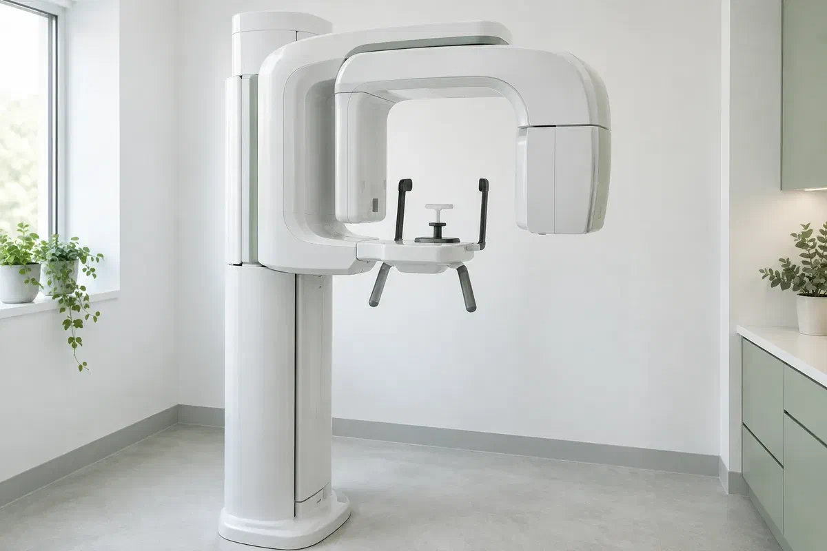
Illustrative discussion near generic panoramic imaging equipment, not actual clinic facilities
General & Family Dentistry

Panoramic Dental X-rays in Burlington

A specialist referral sometimes starts with a request for a wide X-ray of your teeth and jaws. For people coming from Burlington, here is what a panoramic image contributes, how it travels with a referral, and what it leaves for the specialist to assess.

Burlington · referrals and imaging

When another dentist is going to see the picture too.

Suppose your dentist thinks an oral surgeon or orthodontist should weigh in. One of the first things that specialist may want is an overview of your mouth. A panoramic X-ray supplies it: one flat image that takes in the teeth and both jaws together, rather than the close focus of a small film on two or three teeth.

Even with a referral in the works, the image needs its own justification. Your dentist should be able to say what question it answers, how it relates to your examination, and whether a recent X-ray from another office already covers it. A wide view is not something every patient needs just because a file is being opened or updated.

We can talk through whether this kind of image suits your situation and how it would be arranged and shared. Tell us about any recent X-rays, any chance you are pregnant, and anything that makes it hard to stand or keep still. Those details help us plan the imaging and the referral together.

Discuss imaging ahead of a referral
Illustrative unbranded panoramic dental X-ray unit in an empty imaging room
Where the overview tends to help

Position, planning, or a puzzle to solve?

Panoramic imaging fits a particular set of questions about where teeth are and how the jaws appear overall. Your symptoms and the examination show whether it is the right investigation, or whether something else would answer the question more directly.

A tooth that has not erupted normally

A specialist assessing a wisdom tooth or another buried tooth may want to see where it sits in the jaw. The overview can show general position. Ask whether further detail will be needed before any procedure; the image by itself does not decide on extraction.

Treatment covering a wider area

Orthodontic, surgical or restorative planning that involves several teeth can draw on one broad record. It forms part of the planning, alongside other records and the examination. Ask which question it settles and what is left to evaluate.

A finding that raises questions

Swelling, discomfort, an injury or a change in jaw movement may lead to a broader imaging question. Describe your symptoms clearly rather than requesting a particular image. Your dentist should match the investigation to what is going on.

Panoramic images flatten the curve of the jaws onto one plane. Structures overlap, and some shapes are slightly distorted. The image cannot show every fine detail and does not replace a clinical examination, targeted X-rays or other tests where those are needed.

Why referrals often include one

A shared starting point for two clinicians.

With both jaws and the full spread of teeth on a single record, a panoramic image gives the referring dentist and the specialist a common reference. The detector is outside your mouth, which differs from several smaller X-ray techniques. Questions needing finer detail may still call for a different view.

Of course, an image does not interpret itself. Ask which feature relates to your concern, whether anything needs further review, and what the picture cannot tell either clinician. A large image of your mouth should lead to a better explanation, not an assumption that everything has been examined.

Patient information: RadiologyInfo — panoramic dental X-ray

Context around a defined problem

Seeing a single tooth or finding against the rest of the jaw can make a proposed treatment easier to understand. Ask why that context matters in your case. A wider view earns its place when the question needs it.

Records that move with you

Where appropriate, the image and relevant clinical notes can be sent to the specialist. Ask how that is done, how long it may take and whether they will accept it. The specialist will still carry out their own examination.

Clarity about any further test

The overview may answer the question or show why another investigation is needed. Ask what is still uncertain before agreeing to more imaging. A further test should address a specific limitation rather than being ordered automatically.

The imaging appointment

Reason, setup, sweep, review.

Arrangements depend on the equipment and the imaging provider. Ask where the image will be taken and mention anything that may affect your positioning before the appointment is set.

  1. Settle why the image is needed

    Your dentist reviews your history, the examination and relevant earlier records before recommending this view. Ask what it adds for the referral and whether an existing image would be accepted. Mention pregnancy or possible pregnancy so need and precautions can be considered.

  2. Prepare for positioning

    Jewellery, glasses and removable appliances may need to come off. The operator explains the chin rest, bite guide and handholds. Let them know about neck pain, balance problems, restricted movement or difficulty with instructions so adjustments can be made.

  3. Remain still for the rotation

    The imaging arm moves around your head while the picture is captured. Follow the guidance on lip, tongue and head position, and hold steady. If you cannot keep the position, say so before the exposure so the plan can be revisited.

  4. Learn what the image shows

    A clinician has to review the picture and explain it; taking it does not finish the assessment. Ask who will go over the result and when the referral will be sent. If the X-ray was done elsewhere, confirm how it will reach both clinicians.

Between the X-ray and the specialist

Keep the result working for you.

No recovery time or special aftercare is usually needed after a panoramic X-ray. If you had other treatment at the same visit, follow those instructions.

Know how the image affects the plan

Ask whether the result supports the referral, adds a new question or shows nothing further is needed for this issue. Have any technical terms explained. An image is most useful when you understand what it means for the decision ahead.

Keep referral steps moving

Attend the specialist appointment or further assessment when it is offered, and confirm how results will be communicated. Incidental findings should be explained rather than assumed to be harmless or worrying. Ask each clinician what is known and what is not.

Record where the X-ray was taken

Let future dentists and specialists know the date and location of the image so they can decide whether it is still useful. An existing image can sometimes prevent duplication, depending on quality, the question being asked and any changes since.

Imaging choices

Which view does the specialist actually need?

Different methods produce different information. Ask why a particular view is recommended and how its expected benefit weighs against the exposure involved.

For the overall arrangement

A panoramic overview

Useful for selected questions about tooth position and the jaw structures. It uses ionizing radiation and should have a clinical purpose. It is not a stand-in for every other dental view a specialist may want.

For close detail in one area

Targeted intraoral radiographs

A focused image may better answer a question about particular teeth or the bone supporting them. Ask why that level of detail matters. A panoramic image does not automatically remove the need for bitewings or periapical views.

For what the first image left open

Further imaging, if justified

Some referrals lead to another method, such as three-dimensional CBCT where indicated. That is a separate examination, not an automatic next step. The recommendation should name the unanswered question and explain why more information is needed.

Referral and imaging questions

Before the image is sent on.

Imaging decisions should reflect the examination and the information your care needs. Share details of earlier X-rays and raise any questions before the image is taken.

(905) 876-4488
Will the specialist accept a panoramic X-ray taken by my general dentist?
Often, if it is recent, of good quality and answers their question. Some specialists prefer to take their own images or need a different view. Ask both offices before the referral so you are not imaged twice without reason.
How is the image sent to another office?
Digital images are usually transferred securely with your consent, along with relevant notes. Ask how the transfer will happen and whether you should confirm with the specialist that it arrived before your appointment.
Can I get a copy of my panoramic X-ray for my own records?
You can generally request a copy of your dental records, including images. Ask the clinic how to make the request and in what format it will be provided. Keeping a note of where it was taken also helps future dentists.
Does a 2D panoramic tell the surgeon everything a 3D scan would?
No. A panoramic image is two-dimensional. CBCT gives three-dimensional information and has its own indications, and it is not needed in every case. The specialist should explain whether the question requires it.
If I need a panoramic, will my regular bitewings be skipped?
Not necessarily. The wide view shows less detail on some tooth surfaces, so close-up X-rays may still be needed to look for decay. Ask how each image fits into your care.
Who should I tell if I could be pregnant before imaging?
Tell the dentist who recommended the X-ray and the person taking it, before the exposure. They can weigh the clinical need and appropriate precautions with you. Ask how the recommendation applies to you rather than following a general rule found online.
Panoramic X-rays for Burlington referrals

Plan the image and the referral in one conversation.

Contact our Bronte Street practice to discuss your concern, any referral and any imaging that has been suggested. Mention recent X-rays and anything that affects positioning. We will explain the clinical purpose, confirm the available arrangements and show how the result fits into your care.

Imaging is recommended based on clinical need. The equipment photos are illustrative and do not confirm an on-site machine or a specific appointment arrangement.

Visit Milton Dental Clinic
Unit 1, 342 Bronte St. S, Milton, ON L9T 5B7
Get directions Clinic hours
Monday–Thursday
11am–7pm
Friday–Sunday
By appointment only

Areas we serve

  • Milton
  • Oakville
  • Mississauga
  • Georgetown
  • Halton Hills
  • Acton
  • Campbellville
  • Brampton
  • Burlington
  • Puslinch
  • Rockwood
  • Erin
  • Caledon
  • Etobicoke
  • Old Milton
  • Bronte Meadows
  • Timberlea
  • Dorset Park
  • Dempsey
  • Clarke
  • Beaty
  • Coates
  • Harrison
  • Scott
  • Willmott
  • Ford
  • Bowes
  • Cobden
  • Walker
  • Mountain View
  • Milton Heights
  • Hawthorne Village
  • Valley View
  • Fallingbrook
  • Nassagaweya
  • Trafalgar
  • Esquesing
  • Nelson
  • Brookville
  • Moffat
  • Glen Williams
  • Norval
  • Limehouse
  • Stewarttown
  • Ashgrove
  • Hornby
  • Silver Creek
  • Speyside
  • Scotch Block
  • North Oakville
  • River Oaks
  • Glen Abbey
  • West Oak Trails
  • Palermo
  • Bronte Creek
  • College Park
  • Joshua Creek
  • Uptown Core
  • Preserve
  • Iroquois Ridge
  • Lisgar
  • Meadowvale
  • Meadowvale Village
  • Churchill Meadows
  • Erin Mills
  • Central Erin Mills
  • Streetsville
  • East Credit
  • Creditview
  • Heartland
  • Sheridan
  • Mount Pleasant
  • Credit Valley
  • Huttonville
  • Churchville
  • Springbrook
  • Northwest Brampton
  • Bram West
  • Fletcher's Meadow
  • Fletcher's Creek South
  • Alton Village
  • Orchard
  • Millcroft
  • Headon Forest
  • Tansley
  • Brant Hills
  • Tyandaga
  • Kilbride
  • Lowville
  • Aberfoyle
  • Morriston
  • Arkell
  • Hillsburgh
  • Freelton
  • Carlisle
  • Waterdown
  • Flamborough
  • Mountsberg

How can we help?

Milton Dental Clinic

Have a question about your smile? Choose the option that works best for you.

For urgent dental concerns, please call.
Messages are reviewed during clinic hours.