

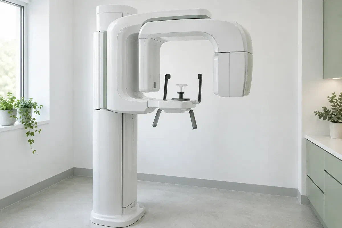
Illustrative discussion near generic panoramic imaging equipment, not actual clinic facilities
General & Family Dentistry

Panoramic Dental X-rays in Caledon

A wide X-ray taken for one reason can show something nobody was looking for. For people coming from Bolton and across Caledon, learn what a panoramic image is meant to answer, how unexpected findings are handled, and what the view cannot confirm.

Caledon · understanding your results

Something else showed up. Now what?

Hearing that an X-ray revealed “something we should keep an eye on” can be unsettling, especially when the image was taken for an unrelated reason. That happens because a panoramic X-ray covers a lot of ground: one flat, two-dimensional picture of the teeth together with both jaws, far wider than the small films used for a few teeth at a time.

Every panoramic should start with a reason. The dentist bases the recommendation on your symptoms, the examination and records already available, rather than on being new or on a set number of years passing. Knowing the original question also helps make sense of any extra finding, because it clarifies what the image was and was not designed to assess.

Our team can discuss whether a wide view suits your situation and how imaging would be arranged. Tell us about earlier X-rays, any chance of pregnancy and anything that makes keeping still a struggle. If a finding comes up, we will explain what it might mean, what is uncertain and what, if anything, should happen next.

Talk through a panoramic result with us
Illustrative unbranded panoramic dental X-ray unit in an empty imaging room
The usual starting points

Most panoramics begin with one of these.

The wide view suits particular questions about where teeth sit and how the jaws look as a whole. Your symptoms and examination decide whether it is appropriate, or whether a narrower or different test would make more sense.

An unerupted tooth

The overview may show the general position of a wisdom tooth or another tooth that has not come through. More detail is sometimes needed before treatment. The image on its own does not decide whether a tooth should be removed, so ask what else will be considered.

Planning across several areas

A broad record may help when treatment covers different parts of the mouth. It complements the examination and other records rather than replacing them. Ask what it will contribute and what will still need assessing separately.

A symptom to investigate

Pain, swelling, an injury or changes in jaw movement may lead to a wider imaging question. Describe what you are experiencing rather than asking for a particular X-ray, so the investigation can be matched to the concern.

A panoramic image flattens curved anatomy, so overlap and some distortion are normal, and shadows can sometimes look like findings. It cannot show every fine detail and does not replace an examination, close-up radiographs or other tests when they are needed.

Why the wide view sees so much

Broad coverage brings answers, and sometimes new questions.

Because it shows both jaws and the full spread of teeth on one record, a panoramic image can reveal features outside the area that prompted it. The detector stays outside your mouth during capture, unlike some smaller dental X-ray methods. Finer detail about any one spot may need another view.

Interpretation is what gives any finding meaning. Ask which part of the picture relates to your original question, whether anything else deserves follow-up, and what the image cannot settle. A large image of the mouth should lead to a careful explanation, not an assumption that all is well or that something serious is present.

Patient information: RadiologyInfo — panoramic dental X-ray

Context for the original question

Seeing a tooth or finding against the whole jaw can make a treatment discussion clearer. Ask how that context applies to you. Wider coverage is useful when the question calls for it, not as a rule.

A record a specialist can review

If a finding needs another opinion, the image and relevant notes may be shared where appropriate. Ask how the referral will work and how long it may take. The specialist will still examine you.

A defined reason for more testing

An incidental finding may need nothing more than monitoring, or it may justify another investigation. Ask what is uncertain and what a further test would clarify before agreeing to it.

From booking to results

How the process runs.

Equipment and procedures depend on the imaging provider. Find out which location will take the image, and mention any positioning concerns before your appointment is finalised.

  1. Name the question up front

    Your dentist considers your history, examination and earlier records before suggesting the view. Ask what the image is meant to answer, and whether a previous X-ray would do. Mention pregnancy or possible pregnancy so need and precautions can be weighed.

  2. Get set at the machine

    Jewellery, glasses and removable appliances may need to come off to avoid shadows. The operator explains how to stand or sit and where to rest your chin. Mention neck pain, balance trouble or limited movement so the setup can be adjusted.

  3. Hold still during the rotation

    The unit moves around your head for a short time. Follow guidance on lip, tongue and head position. If the position cannot be managed, speak up before the exposure so another arrangement can be considered.

  4. Hear about the main and any extra findings

    A clinician needs to review the image and explain it. Ask who will go over it, whether anything unexpected was seen and what happens next. If the X-ray came from a different provider, check how your dentist will get it.

When follow-up is advised

Stay informed, not alarmed.

Panoramic imaging itself needs no recovery period or special aftercare. Instructions for any other treatment done at the same visit still apply.

Ask exactly what was seen

Find out whether the result answers the original question, raises a new one or needs no further action. Ask for any term to be explained in plain words, and whether the finding is common. Understanding it is the first step to deciding what to do.

Follow the monitoring or referral plan

If you are advised to return for review or see a specialist, book it and confirm how results will reach you. An incidental finding should never be assumed unimportant or serious without explanation. Ask what is known and what is still unclear.

Keep the image on record

Tell future dentists when and where it was taken. An earlier image can be valuable for comparison when monitoring a finding, as well as for avoiding unneeded repeats, depending on quality and changes since.

Following up a finding

Sometimes watching is enough; sometimes another view helps.

Different imaging methods show different things. Ask why a particular follow-up view is recommended and what it would add, weighed against the exposure.

To see the broad arrangement

A panoramic overview

Suitable for selected questions about teeth and jaw structures, and sometimes for comparison over time. It uses ionizing radiation, so each image needs a clinical purpose. It does not replace other dental views.

To look closely at one spot

Targeted intraoral radiographs

A small, focused X-ray may clarify a finding near specific teeth or bone. Ask why that detail matters. A panoramic image does not automatically make bitewings or periapical views unnecessary.

To answer what remains unclear

Further imaging if justified

Some findings may warrant another method, such as CBCT, where indicated. It is a separate examination rather than an automatic next step. The dentist should explain the unanswered question and why more detail is needed.

Questions about panoramic results

When the image shows more than expected.

Imaging decisions and follow-up should reflect your examination and care needs. Share details of past X-rays and ask about anything you do not understand.

(905) 876-4488
What is an incidental finding on a dental X-ray?
It is something seen on the image that was not the reason it was taken. Many are harmless variations, but each should be explained. Ask your dentist what it is, how sure they are and whether follow-up is needed.
Who reads my panoramic X-ray?
Your dentist reviews it as part of your assessment. If something falls outside their scope, they may seek a specialist opinion. Ask who is responsible for interpreting your image.
Can an old panoramic be compared with a new one?
Yes, if earlier images are available, comparison can help show whether something has changed. Let your dentist know where previous X-rays were taken so they can be requested.
Should a shadow on the X-ray always lead to more imaging?
Not always. Some shadows are caused by overlap or normal anatomy. The dentist decides based on the finding, your symptoms and the examination. Ask why further imaging is or is not recommended.
Would a Caledon household need to travel for a specialist referral?
That depends on the specialist and where they practise. Ask your dentist about referral options and whether the image can be sent ahead so the first specialist visit is productive.
Can a digital panoramic be taken without radiation?
No. Digital panoramic units still use ionizing radiation, generally at a small dose. Every image should be clinically justified. ‘Digital’ refers to how it is captured and stored.
Panoramic X-rays for Caledon

We’ll explain what the picture means for you.

Reach our Bronte Street practice by phone or online to discuss your concern, any imaging that has been suggested or a finding you would like explained. Mention earlier X-rays and positioning needs. We will go over the purpose, confirm arrangements and show how the result shapes your care.

Imaging is recommended for a clinical reason. Equipment photographs are illustrative and do not confirm an on-site machine or any specific appointment arrangement.

Visit Milton Dental Clinic
Unit 1, 342 Bronte St. S, Milton, ON L9T 5B7
Get directions Clinic hours
Monday–Thursday
11am–7pm
Friday–Sunday
By appointment only

Areas we serve

  • Milton
  • Oakville
  • Mississauga
  • Georgetown
  • Halton Hills
  • Acton
  • Campbellville
  • Brampton
  • Burlington
  • Puslinch
  • Rockwood
  • Erin
  • Caledon
  • Etobicoke
  • Old Milton
  • Bronte Meadows
  • Timberlea
  • Dorset Park
  • Dempsey
  • Clarke
  • Beaty
  • Coates
  • Harrison
  • Scott
  • Willmott
  • Ford
  • Bowes
  • Cobden
  • Walker
  • Mountain View
  • Milton Heights
  • Hawthorne Village
  • Valley View
  • Fallingbrook
  • Nassagaweya
  • Trafalgar
  • Esquesing
  • Nelson
  • Brookville
  • Moffat
  • Glen Williams
  • Norval
  • Limehouse
  • Stewarttown
  • Ashgrove
  • Hornby
  • Silver Creek
  • Speyside
  • Scotch Block
  • North Oakville
  • River Oaks
  • Glen Abbey
  • West Oak Trails
  • Palermo
  • Bronte Creek
  • College Park
  • Joshua Creek
  • Uptown Core
  • Preserve
  • Iroquois Ridge
  • Lisgar
  • Meadowvale
  • Meadowvale Village
  • Churchill Meadows
  • Erin Mills
  • Central Erin Mills
  • Streetsville
  • East Credit
  • Creditview
  • Heartland
  • Sheridan
  • Mount Pleasant
  • Credit Valley
  • Huttonville
  • Churchville
  • Springbrook
  • Northwest Brampton
  • Bram West
  • Fletcher's Meadow
  • Fletcher's Creek South
  • Alton Village
  • Orchard
  • Millcroft
  • Headon Forest
  • Tansley
  • Brant Hills
  • Tyandaga
  • Kilbride
  • Lowville
  • Aberfoyle
  • Morriston
  • Arkell
  • Hillsburgh
  • Freelton
  • Carlisle
  • Waterdown
  • Flamborough
  • Mountsberg

How can we help?

Milton Dental Clinic

Have a question about your smile? Choose the option that works best for you.

For urgent dental concerns, please call.
Messages are reviewed during clinic hours.