Where should the tooth be, and the implant beneath it?
Planning starts from the replacement tooth’s shape, bite and cleaning access. Ask how the implant position on screen was chosen to support that crown rather than the other way round.


Every surgical guide starts as a set of records: a 3D image of the jaw and a scan of your teeth and gum. From Campbellville, find out what those records involve, how they become a guide, how its fit is checked, and why the records alone cannot decide your treatment.
The part of guided implant surgery most people experience first is the record-taking. Usually that means a 3D X-ray of the jaw, which shows bone height, width and nearby structures, plus a digital scan or impression of the teeth and gum. Those files are combined on screen, the implant and future tooth are planned, and a template is designed. That template, the surgical guide, rests on teeth or tissue during surgery and helps direct selected steps.
Good records make planning possible; they do not make the decision. Someone still has to judge whether the bone is adequate, how close the nerve or sinus is, how healthy the gum is, whether instruments can reach, and what the replacement tooth needs. A guide built from excellent scans still cannot make an unsuitable site suitable, and the surgeon’s own assessment continues to matter during the procedure.
When you book from Campbellville, it is worth asking what records you will need and whether any can be taken at a visit you are already making. Then ask which steps the guide would direct, how its fit is tested, and what alternative is planned if it cannot be used. Understanding the chain from scan to surgery is more useful than assuming the screen shows a guaranteed result.
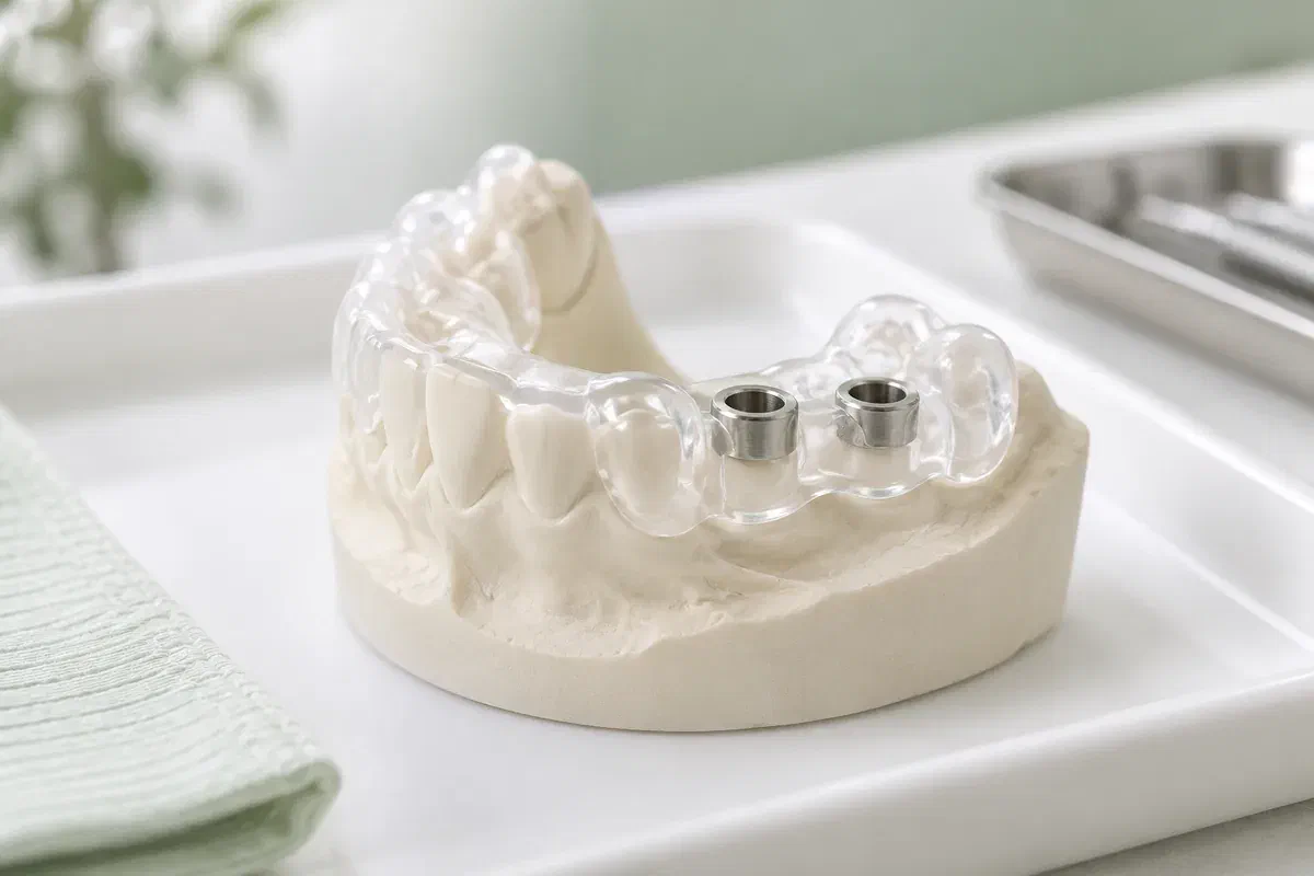
A guide is designed for one specific plan. The conversation should explain what it is meant to achieve and what the records have to show for it to work.
Planning starts from the replacement tooth’s shape, bite and cleaning access. Ask how the implant position on screen was chosen to support that crown rather than the other way round.
Imaging shows bone, nerves, sinus and space for instruments, but every image has limits. The plan still needs a margin for uncertainty and a clinical check of access in the mouth.
Ask which parts of the procedure follow the template and which use other techniques. The answer should include limitations, cost and any extra records or appointments.
A digital plan describes where the implant is intended to go. Guided surgery does not by itself mean no incision, a tooth the same day, no discomfort or faster healing.
If imaging and surface scans are accurately aligned, a guide can translate the planned implant position into a physical template. How well that works depends on the quality of each record, the design and manufacture, the stability of the fit and how the guide is handled.
There is always some gap between planned and achieved positions, and the surgeon must respond to what is actually found. Ask why a guided approach is suggested for you, and how the plan allows for the limits of the records and the guide.
Patient information: International Team for Implantology — computer-guided implant surgery
Combining imaging with a scan of the teeth lets the team see the future crown and the implant together, so you can understand how position affects appearance and cleaning.
Misaligned files can mean a guide that does not fit. Ask how alignment is verified and whether a new filling, a moved tooth or gum change could affect it.
A guide that does not seat properly should not be used just because it was made. Ask how that would be judged and whether the appointment might change.
Workflows differ by plan and guide system. Ask who takes and reviews the records, who designs and produces the guide, who performs surgery and who completes the tooth.
Your dentist looks at the mouth, your health history and the replacement choices, and explains relevant risks. This decides whether records for implant planning are worth taking.
A 3D image and surface scan are combined, the position is planned, and the guide is designed. The team considers its support, access, anatomy, grafting needs and specialist input.
The guide is seated and its fit checked before use. If it does not seat as intended, the surgeon uses an alternative that was already considered rather than forcing it.
Reviews and the timing of the final tooth depend on your healing. Follow-up X-rays may be part of confirming the implant is ready for the restoration.
Instructions depend on what was done, including any extraction, grafting or temporary tooth. Follow the written guidance you receive rather than assuming a guide changes recovery.
Ask how to clean near the implant, which foods to avoid and how to wear any temporary appliance. Comfort early on is not proof that healing is complete.
Keep your instructions and our number handy. Contact us about symptoms they list, unexpected movement, or a problem with a temporary tooth.
Once the crown is placed, cleaning at home and professional visits continue. Periodic X-rays may be used to check the bone around the implant over time.
These answers cover the planning stage in general terms. Ask how each one applies to the records and guide proposed for you.
At our Bronte Street practice, we will examine the site, explain which records would help and discuss whether a guided approach or a specialist opinion makes sense.
If you have had dental X-rays or scans elsewhere recently, bring copies or let us know where they were taken.
